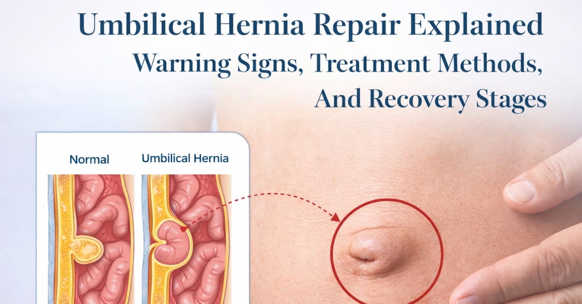
Umbilical hernia repair addresses a bulge close to your navel by closing the defect in the abdominal wall. You get less pain, less risk of bowel complications, and fewer restrictions in daily activities.
Most repairs utilize mesh for added strength, with small incisions and same-day discharge typical. We provide you with specific timelines for rest, wound care, and activity, usually walking the same day.
The following sections discuss treatment options, complications, and recovery.
Key Takeaways
- You can identify an umbilical hernia as a soft bulge at your navel, which can increase in size with coughing, standing, or straining. Go to urgent care if you develop sudden, severe pain, vomiting, or a hard, irreducible bulge.
- You might experience pressure, tenderness, or no pain. Pain that intensifies with lifting or coughing indicates added pressure on the hernia.
- An umbilical hernia occurs when the abdominal wall is weak or damaged, or when it is under repeated pressure. You can reduce risk by maintaining a healthy weight, addressing chronic cough or constipation, and using proper lifting techniques.
- You will likely receive a diagnosis through a physical exam and, if needed, imaging such as an ultrasound or CT to assess size and complications. Early evaluation helps prevent incarceration or strangulation.
- You can opt for watchful waiting for small, asymptomatic hernias, particularly in children. Adults with symptoms or enlarging hernias frequently do well with prompt repair. Talk through your health, lifestyle, and preferences to determine the best timing for surgery.
- You’re looking at open, laparoscopic, or robotic repair that fortifies the abdominal wall. Adhere to post-op directions, avoid strain, and watch for red flags.

What Is An Umbilical Hernia?
An umbilical hernia occurs when the abdominal contents have pushed through the umbilical ring at the belly button. An umbilical hernia is a common abdominal wall hernia in infants and adults. You might notice a bulge that increases with a cough or strain. It can be silent or painful, and when ignored, can lead to complications.
1. The Bulge
Typical symptom is a soft swelling near or through your navel that increases when you stand, lift, or when babies cry. The bulge occurs as intestine or fat protrudes through a defect in the fascia, typically at the attenuated linea alba or former umbilical vessel sites.
It can appear and disappear, is more pronounced with straining, and flattens when you lie down.
Common Signs:
- Size: tiny (≤1 cm) to several centimeters
- Feel: soft, sometimes tender; cough impulse present
- Reducibility: slides back with gentle pressure or when supine
- Changes: enlarges with strain, shrinks at rest
2. The Feeling
You could experience a slight ache, burn, or tenderness at the location. These feelings tend to aggravate with a cough, lifting, or straining because of pressure.
Most hernias are painless and discovered visually only. Acute intense pain with a hard lump that cannot be pushed back in may indicate incarceration or strangulation.
3. The Mechanism
In babies, the umbilical ring sometimes doesn’t shut all the way. In adults, the wall degenerates over time, and a sac containing bowel or omentum protrudes through the fascial defect.
Greater intra-abdominal pressure fuels expansion of the defect. Triggers consist of chronic cough, obesity, pregnancy, ascites, heavy lifting, and longstanding constipation.
Stretching and extra fat can separate muscle fibers and thin aponeuroses, making hernia formation easier.
4. In Infants
Umbilical hernias affect 10% to 15% of newborns, more in premature infants, due to the failure of the ring to close. Most heal on their own by age 2, frequently by age 3 to 4, without treatment.
These are generally painless and low risk. Look out for a hard, non-reducible bulge, pain, or vomiting.
5. In Adults
Adult cases account for 6% to 14% of ventral hernias, second only to inguinal hernias. Risk increases with obesity, multiple pregnancies, previous abdominal surgery, heavy lifting, chronic cough, constipation, and ascites.
While many small hernias, measuring 1 cm or less, are asymptomatic, pain is the number one complaint when present, cited by 44%. As many as 90% of pregnant women will develop one, and repair is delayed unless symptomatic or incarcerated.
Adult hernias almost never resolve and have a higher risk of serious complications, so surgery is recommended if it is painful, growing, or non-reducible.
Why Do Umbilical Hernias Occurs
You get an umbilical hernia when tissue near the navel protrudes through a weak point in the abdominal wall. This abdominal wall defect can exist from birth or develop later as the wall stretches or thins. Understanding umbilical hernia symptoms aids you in planning prevention and selecting umbilical hernia repair techniques that suit your risk profile.
A systematic review conducted in line with guidelines from the College of Surgeons evaluated outcomes of open umbilical hernia repair techniques. When a hernia appears, the contents of the hernia may include fat or bowel, influencing the choice of surgical approach. The review highlighted ongoing debate over mesh versus suture repair, noting that mesh repair generally reduces recurrence rates, particularly in larger defects.
Congenital Weakness
When the umbilical ring fails to close after birth, a gap remains open and creates a route for a hernia. This is the underlying congenital cause. Certain genetic syndromes, such as trisomies and connective tissue disorders, increase risk by altering collagen quality.
The fascia and muscle layers may be thinner or less stiff, so they stretch sooner under average pressure. These defects can affect the linea alba in the vicinity of the navel, where vessels used to traverse generate a navel site of weakness.
Premature babies are at greater risk because their fascia hadn’t fully developed at birth.
Adult Pressures
Repeated strain increases intra-abdominal pressure and eventually expands minor defects. Muscle fibers can separate, and aponeuroses can become attenuated, particularly along the linea alba near the umbilicus. That mechanical creep is why hernias frequently appear after years of slight strain versus a single incident.
Previous abdominal surgery counts. Incisions adjacent to the navel and umbilical-site laparoscopic trocars can create a weaker ring or new defect. Scar tissue cannot bear a load like healthy fascia, so the site bulges with less force.
Up to 90% of adult umbilical hernias are acquired. Risk is more common with obesity, metabolic syndrome, ascites, or a history of multiple pregnancies. Age, injury, and prior surgery contribute to wall weakness.
You reduce risk by addressing constipation, treating cough, using safe lifting techniques, and decreasing central adiposity. Little constant adjustments reduce pressure and preserve the ring’s form and force.

Getting A Diagnosis
You obtain a diagnosis by history, exam, and, when necessary, imaging. Your physician palpates for a navel bulge, inquires about pain, nausea, or vomiting, and screens risk factors such as obesity, advanced age, previous pregnancies, or peritoneal dialysis. Early diagnosis directs safe timing of repair and reduces the risk of incarceration or strangulation.
Imaging, typically an abdominal ultrasound, verifies the defect or alerts to complications. CT is reserved for certain complex situations. Differentiating an umbilical hernia from other abdominal wall masses is important, as lipomas, epigastric hernias, or ascites require distinct management.
Physical Exam
Your physical exam centers on the umbilicus. The clinician looks for a soft or hard bulge that can increase with standing or straining. A noticeable swelling close to the navel is the usual indicator, but certain hernias remain covert.
You might be asked to cough or ‘bear down’ so the hernia protrudes. The examiner then palpates for the defect, determines if it is reducible, and notes size and tenderness. Non-reducibility or sharp pain is concerning for emergent issues.
- Checklist Of Key Findings:
- Location: exact position at or near the umbilicus.
- Size: width of the fascial gap and bulge.
- Reducibility: fully, partly, or not reducible.
- Pain: none, mild tenderness, or severe pain.
- Location: exact position at or near the umbilicus.
Imaging Tests
Imaging clarifies what the sac contains (fat versus bowel), the size of the fascial defect, and any complications of obstruction. It’s useful when the diagnosis is confounding, the hernia is significant or comes back, or symptoms imply a complication.
It distinguishes umbilical hernias from other sources of an abdominal mass.
| Modality | What it shows | When it’s useful | Notes |
| Abdominal ultrasound | Real-time view of the defect and contents | First-line to confirm diagnosis | No radiation, widely available |
| CT scan | Detailed cross-sections, bowel status, complications | Rare, for complex or urgent cases | Helps plan surgery and rule out mimics |
| MRI | Soft-tissue detail without radiation | Selected patients or unclear CT/US | Useful if radiation avoidance is key |
Go to urgent care with severe pain, fever, nausea or vomiting, or unable to pass gas or stool. Some hernias don’t make a sound.
If you have risk factors or a family history, see a clinician even without symptoms. Umbilical hernias can develop at any age, and an early diagnosis stops damage.
The Repair Decision
Balancing watchful waiting versus surgery is based on hernia size, symptoms, and risk of complications. Elective repair is frequently recommended when pain, skin changes, or expansion develops. Your health, goals, and comfort with risk dictate the decision. If surgery is scheduled, the mesh versus suture decision is crucial and must correspond with the defect size and your risk profile.
Watchful Waiting
Watchful waiting means you keep an eye on a small, painless umbilical hernia, but no surgery now. You check for change, keep routine exams, and act if symptoms start. That’s pretty typical for a lot of babies, as pediatric hernias frequently resolve spontaneously by toddler age.
You’ll still need routine follow-up to monitor size in millimetres and new symptoms such as pain, nausea, or skin redness. If symptoms do appear, you switch to repair.
| Situation | Why watchful waiting is not advised |
| Incarceration (bulge won’t reduce) | Trapped tissue risks damage |
| Strangulation (severe pain, red/purple skin, fever, vomiting) | Blood flow cut off; emergency |
Surgical Necessity
You think about surgery if you have pain that restricts activities of daily living, an enlarging defect, or a non-reducible bulge. A number of electrical repairs for timing certainty, and that covers cosmetic concerns if the bulge interferes with clothing or sport.
Technically, the choice between mesh and suture is crucial. Mesh employment is linked to larger abdominal wall hernias, elevated BMI, smoking history, and reoperation. For defects larger than 1 cm and with any risk factor, suture repair yields more recurrences compared to mesh options.
In considering umbilical hernia surgery, one must weigh the benefits of each method. The decision impacts not only the immediate outcomes but also the long-term success rates in preventing hernia recurrence, making preoperative planning essential for optimal results.
Personal Factors
Your age, other illnesses, lifestyle, and job all count. Endurance athletes, manual workers, or those who lift frequently might push you toward earlier repair to avoid flare-ups. Preference matters.
Your tolerance for recurrence risk or for meshes is what guides the plan. Women planning pregnancy and those with heavy-lifting occupations frequently opt for earlier repair to prevent growth and symptoms under strain.
Evidence notes suture repairs have a higher recurrence (about 7.2%) than mesh (about 5.1%), and superficial surgical site infections are linked to recurrence in suture cases. Surgeons may favor mesh for larger defects and suture for small ones.
The final choice depends on defect size and your overall health. Decide on a case-by-case basis with a clear risk review.

Navigating Umbilical Hernia Repair Surgery
You select a technique for umbilical hernia repair depending on the hernia size, your anatomy, and your surgeon’s expertise. All of these choices aim to close the hernia defect and reinforce the abdominal wall. Understanding the compromises allows you to balance comfort, recovery time, and long-term robustness.
An umbilical hernia in adults develops due to weakness in your abdominal wall, allowing tissue from inside your abdomen to protrude through the opening. Unlike hernias in children, hernias in adults often worsen over time and may require surgical intervention rather than observation. During examination, a physician may gently push the hernia back into place, but long-term success depends on treatment. The outcomes of umbilical hernia repair are generally excellent, especially when addressed early and reinforced appropriately to prevent recurrence.
Open Repair
Open repair places the mesh through a single, small cut over the belly button. The sac is released, and the hole is closed with sutures or a mesh if the opening is bigger. Primary suture repair works best when the defect is 2 centimeters or less.
Once it reaches 2 centimeters or larger, most surgeons utilize mesh to reduce tension and decrease recurrence. You can have local or general anesthesia, which is nice if you want to avoid a full anesthetic. Risks include wound infection, hematoma, and a longer recovery than minimally invasive routes.
Expect to stay away from heavy lifts and hard workouts for a couple of weeks.
Laparoscopic Repair
Laparoscopic repair uses 2 to 4 small incisions and a camera to operate inside the abdomen. We use mesh behind the gap to cover and reinforce the hole, which disperses tension and safeguards vulnerable fascia. This is the path that often yields less pain, shorter stays, and a faster return to activities.
It’s a powerhouse choice for bigger or repeat hernias. Imaging with CT or MRI can be used when the exam is unclear to size the defect and inspect its contents. Recurrence after repair runs about 1% to 3% even with mesh.
Robotic Repair
Robotic repair is minimally invasive using wristed tools and HD 3D views. It can assist with tricky cases, like when the umbilical fascia is thin or non-existent, or the round ligament of the liver rests low at the ring.
Precision suturing and mesh placement can improve outcomes for select patients with challenging anatomy. This alternative is typically located at centers with specialized teams.
You still need the basics: stop smoking for 4 weeks, aim for a BMI under 30 kilograms per square meter, and note that nonoperative care is reasonable if you have no symptoms, since the yearly strangulation risk is under 1%.
My Perspective On Recovery
You recover on a schedule dictated by your repair technique (open vs. Laparoscopic, mesh vs. Sutures), the size of your hernia, and your personal health. Adhere to your surgeon’s recovery plan to minimize the chances of infection, seroma, or recurrence.
Step gently back into routine, be on the lookout for red flags, and record shifts.
Ventral hernia repair is commonly performed for umbilical and epigastric hernias when symptoms or complications arise. The treatment of umbilical hernias depends on size and severity, ranging from elective procedures to emergency surgery in cases of incarceration or strangulation. The primary goal is to repair the hernia by restoring the abdominal wall’s integrity, often using sutures or mesh to reduce the risk of recurrence and improve patient outcomes.
The First Week
Anticipate mild to moderate pain, swelling, bruising, and stiffness, particularly in the first 3 to 5 days. Pain and discomfort can persist for as long as two weeks. You can be slower and require assistance getting out of bed or a chair.
Make sure to keep the incision clean and dry as directed. Monitor for spreading redness, warmth, purulent drainage, fever greater than 38 degrees Celsius, increasing pain, or new bulging. These can indicate infection, seroma, or an early recurrence.
Short walks around the home are okay within days. Don’t lift anything heavier than a small bag of groceries. No core work or straining. They’re on pain meds, use them as prescribed on schedule initially, then taper.
Attend your follow-up; they might have to review some stitches or glue, or dressings.
The Long Road
A complete recovery may require weeks to months. If mesh was employed or the hernia was sizeable, anticipate a more gradual ramp-up.
Increase activity in steps: light chores first, desk work when pain is low, then low-impact exercise. Most resume light activities by about 14 days. Heavy lifting and hard workouts typically hold off for 4 to 6 weeks, sometimes more.
Any new or persistent pain, a bulge, tingling, or numb spots require examination. Uncommon nerve or vessel injury may present as changes in sensation or discoloration.
Monitor walking time, pain scores, and sleep. Aim for attainable recovery, not ideal recovery. Scars subside over months; a year or more is typical.
Listening To Your Body When It Happens During An Umbilical Hernia
If pain spikes, redness spreads, fluid accumulates under the skin, or fever increases, notify your care team. A seroma can appear as a soft, squishy lump.
Skip moves that strain your core, such as heavy lifts, sit-ups, and deep twists. Get good sleep, drink water, and consume protein, fiber, and vitamin-rich foods.
Maintain a minimal daily record of steps, pain, meds, bowel movements, and milestones. It assists you and your clinician in observing patterns.

Conclusion
You’re aware of the facts now. About umbilical hernia repair. A defined plan keeps you secure and relaxed. You can balance pain, size, and daily restrictions. You can pose straightforward questions. You can plan your care.
Again, to keep you on track, keep it simple. Record symptoms in a small note on your phone. Schedule consults and follow-ups. Schedule rides, meals, and meds ahead of time. Take light walks to keep the blood flowing. Use a small cough or laugh pillow. Aim for 30 minutes of simple movement a day once cleared. Most return to desk work in 1 to 2 weeks. Most lift light loads by 4 to 6 weeks.
All set for next actions. Book a visit with a board-certified surgeon and bring your list.
FAQ
What is an umbilical hernia?
An umbilical hernia is a protrusion adjacent to your navel. It occurs when tissue bulges through a weak area in your abdominal wall. It may not hurt at all, or it can be painful. Many adults require repair to prevent growth or complications.
How do I know if I need surgery?
You probably require surgical intervention if the hernia becomes larger, painful, or restricts activity. Seek urgent care if it becomes hard, very painful, or if you feel sick. Your surgeon will evaluate the size, symptoms, and risks before recommending repair.
What are my repair options?
Typical choices for umbilical hernia repair include open mesh repair or minimally invasive options like laparoscopic or robotic mesh repair. Using mesh reduces the recurrence risk of abdominal wall hernias. Your selected option depends on hernia size, your health, past operations, and your objectives.
How long is the recovery?
Most individuals return to light activity in 3 to 7 days after umbilical hernia repair. You can return to desk work in 1 week, but no heavy lifting for 4 to 6 weeks. Complete recuperation differs with method and your well-being, so adhere to your surgeon’s instructions.
What risks should I consider?
Risks include infection, bleeding, pain, seroma, mesh problems, and recurrence. With an experienced surgeon, serious complications are rare. Quitting smoking, controlling weight, and managing diabetes lowers risks. Discuss your own risk profile prior to surgery.
How do I prepare for surgery?
Quit smoking, control your weight, manage your blood sugar, and check your medications. Schedule a ride and assistance at home. Follow fasting orders. Query your surgeon about pain control, mesh type for hernia repair, and aftercare. Preparation makes outcomes better and recovery faster.
What does recovery feel like?
Anticipate soreness around the navel, some swelling, and tiredness after umbilical hernia surgery. Early walking is beneficial for recovery. Use your pain plans and support your abdomen when you cough. Call your surgeon for fever, increasing pain, redness, or vomiting. Most patients improve every day.

 Alana3536 says:
 Brooke986 says:
 Anastasia1913 says:
 backlinks says:
 Dale4189 says: